

🎓
Download your CPD certificate
Only participants who attended a webinar in full are eligible for the certificate.
🕐 To convert webinar times to your local time zone, use the Greenwich Mean Time converter.
🎓 For certificate queries, visit the certificates page.
📢 IOMP School Webinars run approximately monthly — watch this page and IOMP social media for upcoming announcements.
▶ Upcoming
No upcoming webinars scheduled at the moment — please check back soon!
In the meantime, browse our past webinar recordings below.
Past webinars 2026 — recordings available
Date & time
Wed 8 July 2026 · 12:00–13:00 GMT
Duration
1 hour
CPD / CE
1 point
Organizer
Eva Bezak, IOMP President
Moderator
Francis Hasford, IOMP Publication Committee Chair
Bringing MRgART into the Clinic — Why and How?
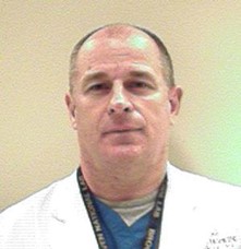
Dr. Simon Woodings, PhD — Medical Physicist, Department of Radiotherapy, University Medical Center Utrecht, The Netherlands
▼ Abstract
Magnetic resonance image guided online adaptive radiotherapy (MRgART) is enabling a new vision of the patient and new radiotherapy treatments at many centres throughout the world. It is an exciting element of the broader sweep toward online adaptive radiotherapy (oART) based upon CBCT, CT, PET or MR imaging. Radiation oncologists wish to use this modality, especially for treating soft-tissue targets in the abdomen and pelvis, where (i) soft-tissue contrast is necessary and (ii) targets and organs-at-risk deform and move, including during treatment. Commissioning and clinical implementation include some new physics concepts and additional checks not needed in conventional radiotherapy. These will be discussed along with some QA results and tips.
▼ Speaker biography
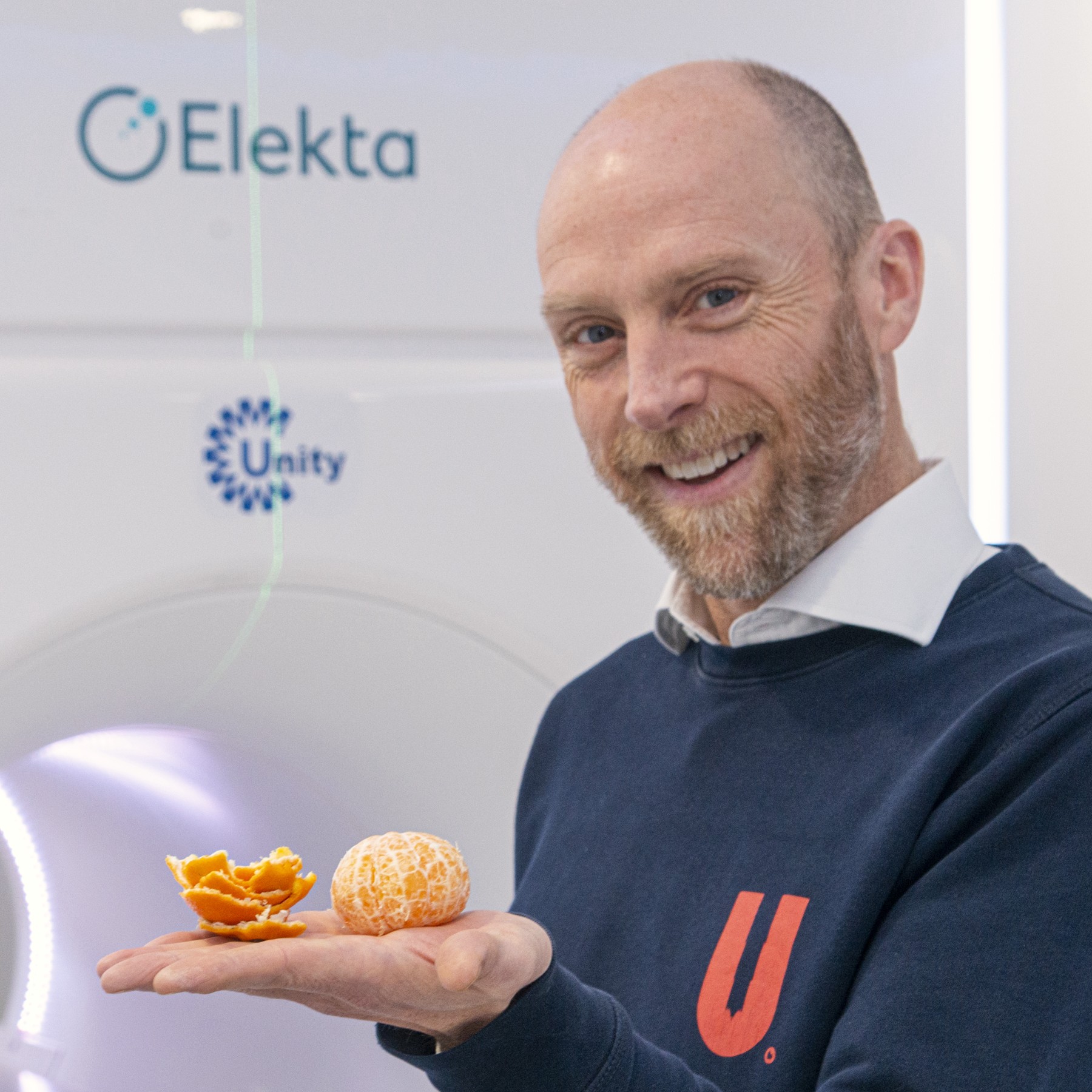
Dr. Simon Woodings, PhD is a radiotherapy medical physicist at UMC Utrecht, the Netherlands, where, over the last 11 years, he has been part of the team contributing to the development, clinical implementation, and clinical use of the Unity 1.5T MR-Linac. He is registered for clinical practice in Australia and the Netherlands and was previously an examiner for the Australasian College of Physical Scientists and Engineers in Medicine (ACPSEM). He coordinates the Utrecht International MRinRT course and is developing MR-Linac QA guidelines with the American Association of Physicists in Medicine (AAPM TG352).
By the end of this webinar, you will be able to:
- Describe some of the oART systems in the world and some of their clinical advantages.
- Name one or more MRgART references that should be consulted prior to clinical implementation and use.
- Describe several limitations of MRgART systems.
- List three or more new critical elements and tests that apply to the Unity 1.5T MR-Linac.
Date & time
Wed 10 June 2026 · 12:00 GMT
Duration
1 hour
CPD / CE
1 point
Organizer
Mika Kortesniemi, PhD — IOMP ETC Chair
Moderator
Jaydev K. Dave, PhD, DABR, MS, FAAPM
3S for MRI — Scanners, Sequences and Safety
Dr. Eric G. Stinson, PhD, MS — MR Medical Physicist, Mayo Clinic, Rochester, MN, USA
▼ Abstract
This lecture provides a comprehensive overview of key concepts in Magnetic Resonance Imaging (MRI), focusing on the technology, image formation, and safety principles critical to clinical practice. We will explore the wide range of MRI scanner field strengths — from low-field to ultra-high-field systems — highlighting their respective advantages, limitations, and clinical applications. The session then introduces the major categories of pulse sequences, including spin echo, gradient echo, and advanced fast-imaging techniques, emphasizing how sequence design influences tissue contrast and diagnostic utility. Finally, we will review key administrative controls that support MR safety, including screening procedures, zoning, staff training, and incident-prevention strategies. By the end of this lecture, participants will gain a clearer understanding of how scanner characteristics, sequence selection, and robust safety practices work together to ensure high-quality and safe MRI examinations.
▼ Speaker biography
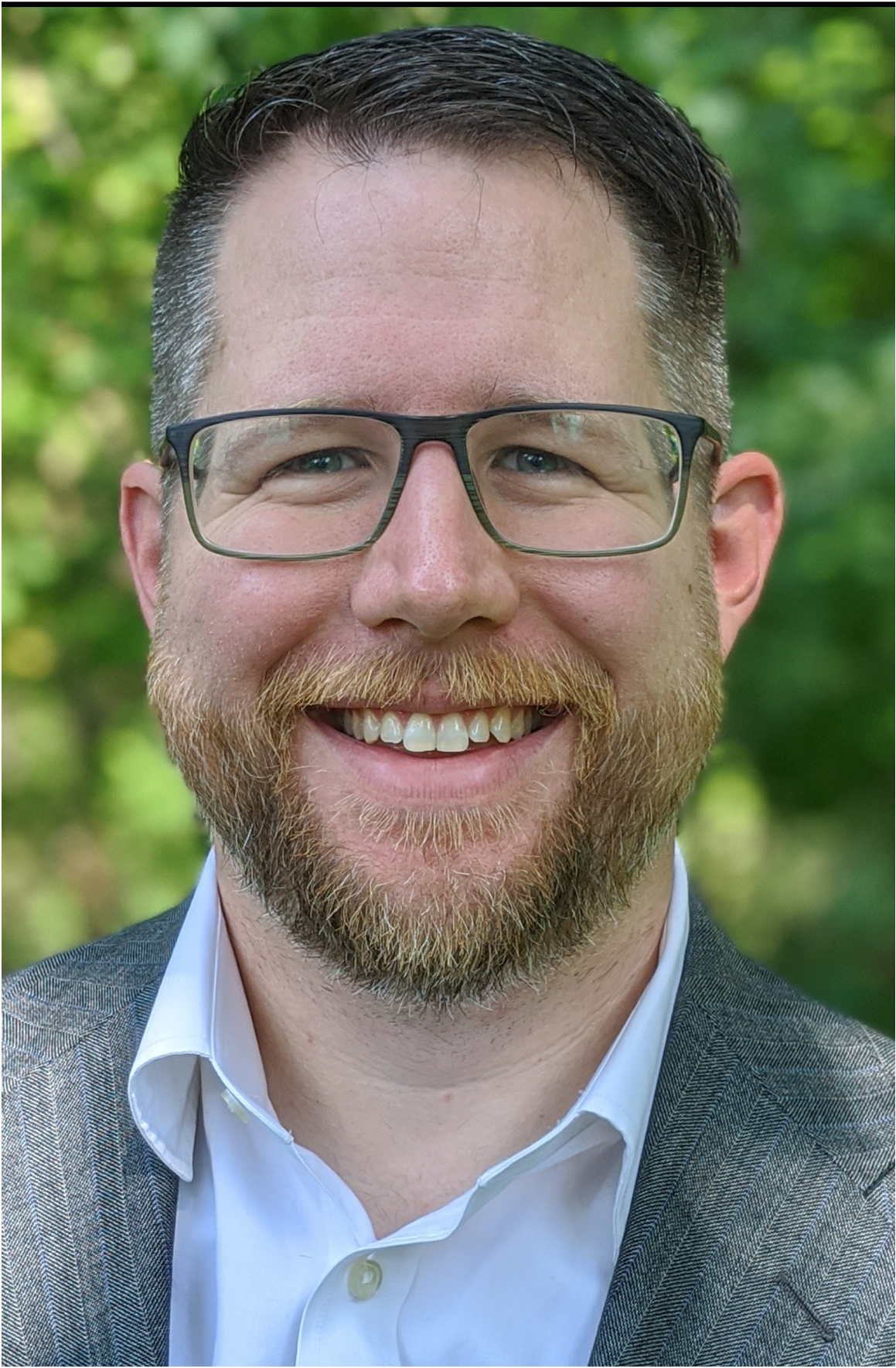
Dr. Eric G. Stinson, PhD, MS is an ABMP-certified MRI medical physicist at the Mayo Clinic in Rochester, MN, USA. He received his master’s degree in medical physics from McGill (Montreal, QC, Canada), working with Dr. G. Bruce Pike at the Brain Imaging Centre at the Montreal Neurological Institute. He then pursued a PhD in biomedical engineering and physiology at Mayo Clinic, working with Dr. Stephen Riederer on improving vessel lumen depiction in contrast-enhanced MR angiography through advanced image reconstruction techniques. Dr. Stinson subsequently worked in ultra-high field MRI for Siemens Healthineers for 4 years before returning to Mayo Clinic as a clinical MR medical physicist. Throughout his career he has been involved in RF coil design, diffusion imaging, highly accelerated time-resolved contrast-enhanced MR angiography, perfusion imaging, fat-water separated imaging, and ultra-high-field MR imaging. He lectures in the MRI technologist programme at Mayo Clinic School of Health Sciences and is a past organizer of the ISMRM Artifact Game Show.
Learning objectives:
- Scanners: Identify the wide variety of field strengths in MRI and the advantages and disadvantages of each.
- Sequences: Understand the main types of pulse sequences and how they are used to produce differing contrast.
- Safety: Identify the administrative controls used to ensure MR safety.
May
20
2026
⌄
May
20
2026
Date & time
Wed 20 May 2026 · 12:00 GMT
Duration
1 hour
CPD / CE
1 point
Organizers
Magdalena Stoeva & Eva Bezak
Moderator
Eva Bezak, IOMP President
Talk 1: From Commissioning to Clinical Use of the LAP LUNA 3D SGRT System
Dr. Hui Khee Looe — Deputy Head of Medical Physics, Pius-Hospital, Oldenburg, Germany
▼ Abstract
Surface Guided Radiation Therapy (SGRT) has become an important component of modern radiotherapy. The LUNA 3D SGRT system enables precise patient positioning for each treatment fraction and supports reliable motion monitoring during treatment. Its comprehensive reporting capabilities allow traceable records of patient positioning and motion throughout the treatment course.
This presentation outlines the testing, implementation, and clinical evaluation of LAP’s LUNA 3D SGRT installed on C-arm linacs. Acceptance testing was performed in accordance with the ESTRO-ACROP guideline, including End-to-end testing encompassing a laser-free workflow. Post-clinical implementation evaluation was performed, including a comparison of patient positioning achieved with LUNA 3D and a conventional laser-based setup. All test results were within manufacturer’s specifications and ESTRO guideline tolerances.
This presentation outlines the testing, implementation, and clinical evaluation of LAP’s LUNA 3D SGRT installed on C-arm linacs. Acceptance testing was performed in accordance with the ESTRO-ACROP guideline, including End-to-end testing encompassing a laser-free workflow. Post-clinical implementation evaluation was performed, including a comparison of patient positioning achieved with LUNA 3D and a conventional laser-based setup. All test results were within manufacturer’s specifications and ESTRO guideline tolerances.
▼ Speaker biography
Dr. Hui Khee Looe is the Deputy Head of Medical Physics at Pius-Hospital in Oldenburg, Germany, and a scientist in the “Medical Radiation Physics” group at the University of Oldenburg. He leads a research group focusing on mathematical and computational methods in dosimetry, and works actively with the clinical team to improve clinical workflow and patient outcomes using the LUNA 3D SGRT system.
Talk 2: Open-Source Tools for the Clinic – Browser Based Tools
Ryan Clark — Senior Research Scientist, Medical Affairs, Siemens Healthineers
▼ Abstract
This session presents five browser-based, open-source tools developed within the Clinical Physics Innovations team at Siemens Healthineers. The tools cover dose constraint conversion/template creation, dosimetric scorecard evaluation, plan delivery visualization, SFRT sphere lattice generation and virtual cone planning. Each runs entirely in a modern browser with no installation required and no patient data leaving the local machine.
▼ Speaker biography
Ryan Clark is a Senior Research Scientist in Medical Affairs at Siemens Healthineers, connecting clinical practice, research and product development to improve workflows for radiation therapy professionals. He also develops open-source radiation therapy software using AI to translate clinical knowledge directly into working software.
Learning objectives:
- Identify browser-based open-source tools designed to address common workflow gaps in radiation therapy planning.
- Describe how AI-assisted development can enable clinicians to build and share practical tools with the broader radiation therapy community.
Apr
20
2026
⌄
Apr
20
2026
Date & time
Mon 20 April 2026 · 12:00 GMT
Duration
1 hour
CPD / CE
1 point
Organizer
Eva Bezak, IOMP President
Moderator
Eva Bezak, IOMP President
Part 1: Delivering Sustainable Healthcare through Collaboration
Fiona Adshead, PhD — Chair, Sustainable Healthcare Coalition
▼ Abstract
This talk addresses why now is the time for action on climate and health, and how by measuring and understanding our impact we can be sustainable by design. By exploring how climate and health are linked, the talk looks at impact across the healthcare value chain, the emerging models of sustainable healthcare, and the need to transform clinical delivery through technology and innovation. Using kidney care as an example, the talk describes how clinicians are driving change through novel approaches to procurement and a re-examination of how to measure value in healthcare.
▼ Speaker biography
Fiona Adshead chairs the Sustainable Healthcare Coalition. Her previous roles include Deputy Chief Medical Officer and Director General in the UK Government, Director of Chronic Disease and Health Promotion at the World Health Organisation, and Bupa’s Chief Wellbeing Officer. She is a Visiting Professor at UCL and Senior Associate at the Cambridge Institute for Sustainability Leadership.
Part 2: Environmental sustainability in medical physics: what we can do
Dr. Robert Chuter, PhD — Principal Clinical Scientist (Radiotherapy), The Christie NHS Foundation Trust & University of Manchester
▼ Abstract
Healthcare currently contributes 4–5% of global greenhouse gas emissions worldwide, with oncology patients being particularly vulnerable to the climate crisis. As scientists we are well placed to estimate the environmental impact of our services through measuring our carbon footprint and performing Life Cycle Analyses. These can help identify the carbon hot spots in our services and highlight where to focus efforts to reduce our carbon footprints. Practical steps will be covered as well as how to build your own sustainability group.
▼ Speaker biography
Dr. Robert Chuter works in a translational role at The Christie NHS Foundation Trust and the University of Manchester, focusing on the MR-Linac. He founded IPEM’s environmental sustainability group in February 2020, leads a joint working party with the RCR and SCoR, and is co-lead for the Green Network within ESTRO.
Apr
21
2026
⌄
Apr
21
2026
Date & time
Tue 21 April 2026 · 12:00 GMT
Duration
1 hour
CPD / CE
1 point
Organizer
M. Mahesh, IOMP Vice President
Moderator
M. Mahesh, IOMP Vice President
The Role of Medical Physicists in Promoting More Environmentally Sustainable Practices in Medical Imaging
Diana Carver, PhD (Vanderbilt University Medical Center) & Andrew M. Hernandez, PhD (UC Davis Health)
▼ Abstract
Medical physicists are central to the safe and effective use of radiation, ensuring imaging systems deliver diagnostic-quality images and supporting the implementation of emerging technologies. This webinar highlights practical opportunities for medical physicists to drive meaningful, sustainability-focused change in clinical imaging practice, including advancing energy-efficient scanner design, minimizing medical waste, optimizing clinical workflows, and reducing unnecessary imaging.
▼ Speaker biography
Diana Carver, PhD is an Associate Professor of Clinical Radiology and Radiological Sciences and a diagnostic imaging medical physicist at Vanderbilt University Medical Center. She serves as Vice-Chair of the AAPM Working Group on Environmental Sustainability in Medical Imaging Physics.
Andrew M. Hernandez, PhD is an Associate Professor of Radiology in Medical Physics and Director of Sustainability at UC Davis Health. He is Chair of the AAPM Working Group on Environmental Sustainability in Medical Imaging Physics and a member of the RSNA Environmental Sustainability Committee.
Apr
22
2026
⌄
Apr
22
2026
Date & time
Wed 22 April 2026 · 12:00 GMT
Duration
1 hour
CPD / CE
1 point
Organizer
Eva Bezak
Moderators
Chai Hong Yeong & Kwan Hoong Ng
Healthcare Sustainability in LMIC Countries
Hasin Anupama Azhari (Bangladesh) · Patricia Mora Rodríguez (Costa Rica) · Iyobosa B. Uwadiae (Nigeria)
▼ Abstract
This webinar presents perspectives from three IOMP regions — Latin America, Africa, and Asia — on the role of medical physicists in supporting more sustainable healthcare systems in low- and middle-income countries (LMICs). Through regional experiences and examples, the session highlights how medical physicists contribute to sustainability by adapting practices to local clinical, economic, and infrastructural contexts, emphasizing collaboration, innovation, and professional responsibility in advancing sustainable healthcare delivery.
▼ Speaker biography
Professor Hasin Anupama Azhari is a pioneering medical physicist in Bangladesh, internationally recognized for advancing cancer care, education, and professional development. She is the current President of AFOMP and Honorary Director of the South Asia Centre for Medical Physics and Cancer Research (SCMPCR). Honored with the IOMP International Medical Physics Award (2018) and AFOMP Outstanding Medical Physicist Award (2020).
Patricia Mora Rodríguez is a medical physicist with over 40 years of experience in medical imaging and radiation protection. She is a former professor at the University of Costa Rica and served as President of ALFIM (2022–2025). Her professional focus includes quality assurance, radiation dose optimization, and capacity building in low- and middle-income countries.
Iyobosa B. Uwadiae is the President of the Nigerian Association of Medical Physicists (NAMP), Secretary of the NAMP Clinical Training and Certification Board, Chair of the Professional Development Committee of FAMPO, and a member of the IOMP Women’s Sub-Committee. She is Board Certified by the International Medical Physics Certification Board (IMPCB).
Apr
23
2026
⌄
Apr
23
2026
Date & time
Thu 23 April 2026 · 12:00 GMT
Duration
1 hour
CPD / CE
1 point
Organizers
Mohammad Hassan Kharita & Mika Kortesniemi
Moderators
Mohammad Hassan Kharita & Mika Kortesniemi
Remote and Automated QC in Radiology – an IAEA Approach
Virginia Tsapaki (IAEA) · Mohammad Hassan Kharita, PhD (Hamad Medical Corporation) · Mika Kortesniemi, PhD (University of Helsinki)
▼ Abstract
This session introduces the possibilities and benefits of technical image QA analysis software facilitating automated image QA in remote regions with limited medical physicist resources. The presented tools are part of the IAEA Coordinated Research Project “Advanced Tools for Quality and Dosimetry of Digital Imaging in Radiology” (E24025). The methodology utilises simple test objects, automated analysis software, and networked data sharing — enabling more frequent quality monitoring while reducing workload, expert time, and travel costs.
▼ Speaker biography
Dr. Virginia Tsapaki is a Technical Officer in the IAEA DMRP Section with over 34 years of experience in medical imaging, quality and safety. She has coordinated more than 120 IAEA technical-cooperation projects and co-authored five IAEA guidance documents working with more than 90 expert contributors.
Dr. Mohammad Hassan Kharita is Acting Executive Director of Medical Physics at Hamad Medical Corporation (HMC), overseeing all medical physics operations across 16 hospitals. He serves as IOMP Treasurer and MEFOMP President for 2025–2028.
Dr. Mika Kortesniemi is Chief Medical Physicist and Adjunct Professor at the HUS Diagnostic Center, University of Helsinki and Helsinki University Hospital. His research focuses on quality assurance, radiation dosimetry, optimisation and radiation protection in x-ray imaging, especially CT and AI.
Apr
24
2026
⌄
Apr
24
2026
Date & time
Fri 24 April 2026 · 12:00 GMT
Duration
1 hour
CPD / CE
1 point
Organizer
Magdalena Stoeva
Moderator
Eva Bezak
SHAPES: Sustainable Healthcare through Advancement of Physical and Engineering Sciences
Ratko Magjarević (IUPESM President) · Marcia Barbosa (ISC Vice-President) · John Damilakis (IUPESM Vice President)
▼ Abstract
This webinar explores how the collaboration between medical physicists and biomedical engineers shapes healthcare’s technological landscape. Enhancing healthcare through capacity building, networking, R&D, and combined efforts of global and local players will contribute to a sustainable environment. The SHAPES webinar is part of the IUPESM MEP initiative, a platform for collaboration between medical physicists and biomedical engineers.
▼ Speaker biography

Ratko Magjarević received his PhD in Electrical Engineering from the University of Zagreb and is a full professor in Electronic Instrumentation and Biomedical Engineering. He was elected President of IFMBE from 2022–2025 and is currently IUPESM President 2025–2028.
Marcia Barbosa is full professor at Universidade Federal do Rio Grande do Sul, a member of the Brazilian Academy of Sciences and the World Academy of Sciences, and Rector of UFRGS since 2024. She is Vice-President of the International Science Council since 2025.
John Damilakis is a full professor and Chairman of the Department of Medical Physics at the University of Crete. He is Immediate Past President of IOMP and Vice President of IUPESM. He is recognised in Stanford University’s list of the “World’s Top 2% Scientists” with 280 PubMed-indexed articles and an h-index of 57.
Mar
6
2026
⌄
Mar
6
2026
Date & time
Fri 6 March 2026 · 12:00 GMT
Duration
1 hour
CPD / CE
1 point
Organizer
Loredana Marcu, IOMP Women Sub-Committee Chair
Moderator
Loredana Marcu, IOMP Women Sub-Committee Chair
Talk 1: The impact of Women in Physics on IUPAP and its mission to assist in the worldwide development of physics
Silvina Ponce Dawson, PhD — FCEN-UBA / CONICET, Argentina · President, IUPAP
▼ Abstract
This talk presents a journey within IUPAP, starting as a member and Chair of the Working Group on Women in Physics and eventually leading to becoming the second woman President of IUPAP in its 104 years of existence. It shows how the inclusion of a gender perspective in scientific institutions can lead to better processes and increase their impact.
▼ Speaker biography
Silvina Ponce Dawson is Full Professor at the University of Buenos Aires, Higher Researcher at CONICET, and the second woman President of IUPAP. Her main research interests are in biological physics and nonlinear dynamics. She has authored over 100 scientific papers and advised 28 theses.
Talk 2: Leading as a woman: Resilience and the power of Perspective
Robin Miller, MS, DABR, FAAPM — President of AAPM · Chief Medical Physicist, Kaiser Permanente Capitol Hill
▼ Abstract
This presentation highlights the resilience and strategic skills women develop in leadership roles — thorough preparation, reflective decision-making, and careful communication. It explores how these adaptive behaviors reflect dedication to fostering trust and achieving results, and how recognizing these strengths shifts the narrative toward equity and confident, authentic leadership.
▼ Speaker biography
Robin Miller is a nationally recognized leader in radiation oncology physics with over three decades of experience. She is the sixth woman to be President of AAPM and has served twice on the AAPM Board of Directors. She is a Fellow of AAPM and board-certified by the American Board of Radiology.
Talk 3: My Career, My Way — A Woman for All Seasons
A/Prof Natalka Suchowerska, PhD, FACPSEM, MESTRO — University of Sydney · Chair CEAB of IOMP
▼ Abstract
This personal reflection challenges the idea of a single “correct” path to success in medical physics, addressing challenges such as imposter syndrome, underrepresentation, and work–life integration, each paired with practical strategies and lessons learned. Aimed at students, early-career professionals, and established physicists alike.
▼ Speaker biography
A/Prof Natalka Suchowerska has worked at the intersection of research, clinical practice, and academia for nearly four decades. She is a founding member of the Help Ukraine Group (HUG), a NAATI-certified Ukrainian–English interpreter, and was recognized in the AFR Top 100 Women of Influence (Innovation) and ESTRO Honorary Membership (2021).
Feb
9
2026
⌄
Feb
9
2026
Date & time
Mon 9 February 2026 · 12:00 GMT
Duration
1 hour
CPD / CE
1 point
Organizer
M. Mahesh, IOMP Vice President
Moderator
M. Mahesh, IOMP Vice President
Talk 1: Introduction to RPT Dosimetry
Robert F. Hobbs, PhD — Dept. of Radiation Oncology and Molecular Radiation Sciences, Johns Hopkins University School of Medicine
▼ Abstract
Radiopharmaceutical Therapy (RPT) is rapidly becoming a mainstream modality. Unlike other systemic cancer treatments, RPT is radiation-based, allowing quantification of activity distribution and conversion to absorbed dose. This talk presents the basic principles of RPT dosimetry and basic QA methods, highlighting how personalized dosimetry-based treatment planning could dramatically improve efficacy of treatments and patient outcomes.
▼ Speaker biography
Robert F. Hobbs, PhD is an Associate Professor and Medical Physicist in Radiation Oncology at Johns Hopkins. He is Chairman of the AAPM Radiopharmaceutical Therapy sub-Committee and recipient of the 2025 MIRD Loevinger-Berman award.
Talk 2: An Introduction to Theranostics
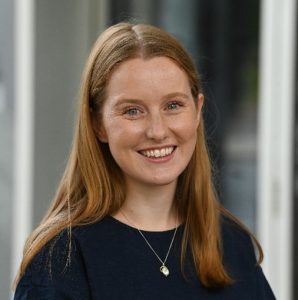
Ashleigh Hull, PhD — School of Allied Health & Human Performance, Adelaide University, South Australia
▼ Abstract
Theranostics represents a rapidly expanding paradigm in nuclear medicine, combining diagnostic imaging and targeted radionuclide therapy to enable personalised patient care. This talk introduces the fundamental principles of theranostics, including theranostic targets, radionuclide selection, imaging–therapy pairs, and clinical examples in practice.
▼ Speaker biography
Ashleigh Hull, PhD is a Lecturer in Nuclear Medicine at Adelaide University and a registered nuclear medicine technologist. Her PhD focused on the pre-clinical development of novel radioimmunoconjugates for the treatment of pancreatic cancer, enabling her to observe the full translational pathway from bench to bedside.
Jan
20
2026
⌄
Jan
20
2026
Date & time
Tue 20 January 2026 · 12:00 GMT
Duration
1 hour
CPD / CE
1–2 CE points
Organizer
—
Moderator
Carmel J. Caruana, PhD — University of Malta
CE credit: 2 CE credit points for participants who complete and pass the associated assessment · 1 CE credit point for other participants
An Introduction to Data Analysis with Python for Medical Physics
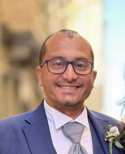
Eric Pace, M.Sc., MIPEM, MPE, RPE — Medical Physics, University of Malta
▼ Abstract
Python has become increasingly prominent in scientific and clinical computing, including Medical Physics and AI applications. This webinar provides a practical overview of fundamental topics including data analytics using pandas and matplotlib, reading DICOM files, and analysing Catphan QC images using pydicom and pylinac.
The 6th edition of the EBAMP-accredited Data Analysis with Python for Medical Physics course (endorsed by EFOMP and IOMP) is planned for 29–31 October 2026 — see thepythoncourse.eu.
The 6th edition of the EBAMP-accredited Data Analysis with Python for Medical Physics course (endorsed by EFOMP and IOMP) is planned for 29–31 October 2026 — see thepythoncourse.eu.
▼ Speaker biography
Eric Pace is a medical physics and radiation protection expert in diagnostic and interventional radiology with over ten years of clinical experience. He lectures in Diagnostic and Interventional Radiology and Python for Medical Physics at the University of Malta, and is President of the Malta Association of Medical Physics.