IOMP SCHOOL WEBINARS 2023

Past Webinars 2023
19 Dec 2023: IOMP Webinar: IOMP’s Focus: Early Career in Medical Physics and the most recent trends in diagnostic imaging and radiotherapy
View Recording
19 December 2023 at 12 pm GMT; Duration 1 hour
Organizer: M Stoeva
Moderator: KH Ng
Speakers: Tan Hong Qi & Choirul Anam, IUPAP Early Career in Medical Physics awardees
Title: Impact of dispersive proton beam on reference dosimetry in a synchrotron spot scanning system

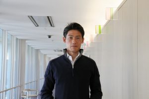
Dr Tan Hong Qi is currently a senior medical physicist in National Cancer Centre of Singapore (NCCS) and is also a clinical instructor in Duke-NUS medical School. He received his PhD from National University of Singapore and completed his residency in NCCS. He is active in both the radiotherapy research and education in Singapore for which he was recognized with institutional level awards and recent SEAFOMP young leader award. His current research interests are the application of AI in radiotherapy, adaptive radiotherapy, and proton therapy.
Abstract:
We have recently commissioned the largest proton therapy center in Singapore and have treated more than 30 patients over the last 4 months after the first clinical start date. This talk will share some of our commissioning experience with Hitachi ProBeat proton therapy which is a spot scanning synchrotron delivery system. In particular, we will report the observation of a large fluctuation in reference dosimetry measurement with a square field and an ion chamber measurement at the plateau region of a monoenergetic proton beam. An investigative journey reveal that the fluctuation originated from the dispersion in the proton beam. This accelerator physics term will be introduced, and we will show for the first time, the feasibility of measuring it in the clinic.
Title: Automated dose and image quality measurements from clinical CT images for CT optimization
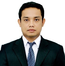
Dr. Choirul Anam completed his Ph.D at Physics Department, Bandung Institute of Technology (ITB). He received Master degree from University of Indonesia (UI) and the B.Sc degree in physics from Diponegoro University (UNDIP). He is currently working as a Lecturer and Researcher in the UNDIP. His research interests are medical image processing and dosimetry for diagnostic radiology, particularly in CT. He has authored and co-authored over 200 papers. In the last ten years, he was the first author over 50 papers. One of his papers published in the Journal of Applied Clinical Medical Physics had been awarded as the Best Medical Imaging Physics Article in 2016. He received the Best Paper Award during 15th South East Asian Congress of Medical Physics (SEACOMP). He was also recognised as an Outstanding Reviewer for Physics in Medicine and Biology in 2018 and for Biomedical Physics and Engineering Express in 2019. He was also awarded as the SEAFOMP Young Leader Awards 2019 for his contribution in CT dosimetry and image quality from the South East Asian Federation of Organizations for Medical Physics (SEAFOMP). He is developer of two software, i.e. IndoseCT and IndoQCT (www.indosect.com).
Abstract:
Recent studies revealed that computed tomography (CT) scanning poses a potential cancer risk in patients. Although the risk is considered much smaller than the benefits of CT scanning to accurately diagnose patient abnormalities, the CT examination must be optimized. In radiation optimization, an image of sufficient quality to diagnose the patient must be achieved with the smallest possible dose. Up until now, image quality and radiation dose were usually evaluated using a standard phantom of standardized characteristic. The evaluation of image quality and radiation dose using the standard phantom is very good for assessing dependency on controllable variables, such as tube currents, tube voltage, pitch, reconstruction filter, and beam width. However, they are also influenced by uncontrollable variables, such as body size and body region. For optimization purposes, the controllable and uncontrollable variables must be considered simultaneously. This talk will discuss methods of automated image quality (noise and spatial resolution) and patient dose (i.e. size-specific dose estimate (SSDE)) directly from image of patient.
7 Nov 2023: IOMP Anniversary Webinar: November 07, 2023
Moderators: John Damilakis & Eva Bezak
Speakers: Azam Niroomand-Rad, Colin G. Orton, Fridtjof Nüsslin
Title: The 60th Anniversary of IOMP – Personal Memories and Some Thoughts on the Future of Medical Physics
Speaker: Prof. Azam Niroomand-Rad, PhD, DSc, FAAPM, FACMP, DABR, FIOMP, FIUPESM
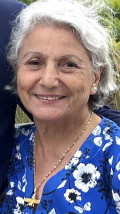
Azam Niroomand-Rad was born in Tehran, Iran. Encouraged by her math/physics teacher and inspired by Maria Skłodowska-Curie, she won Fulbright Scholarship for college education and went to USA in 1966. Azam completed BS in Math/Physics with honors (Summa Cum Laude & Phi Beta Kappa) from State University of NY in 1970 and then PhD in Atomic and Molecular Physics from Michigan State University in 1978.
Azam Niroomand-Rad received Postdoc Fellowship in 1981 and worked at the Department of Medical Physics at University of Wisconsin in Madison, Wisconsin under supervision of late Profs John Cameron and Herb Attix. She has specialized in therapeutic medical physics and has been certified by American College of Radiology (ACR) in 1988 by American Board of Medical Physics (ABMP) in 1990.
Prof. Niroomand-Rad worked at Medical College of Wisconsin in Milwaukee (1983-1988) and then at the Department of Radiation Medicine, Georgetown University Medical Center, Washington DC until her retirement in 2008. She is now an Honorary Prof. at University of Wisconsin-Madison where she lives.
Prof Azam Niroomand-Rad is first (and thus far only) woman to serve as IOMP President (2003-2006). She is founder of the AAPM / IOMP International Scientific Exchange Programs (ISEP) for developing countries and has served on many AAPM / IOMP committees / Task Forces including International Labor Organization (ILO) (1991- 2008). She has published numerous articles and book chapters and has been Co-Inventor of a US Patent for designing a novel stereotactic method for treatment of spine lesions. She has received many Honors and Awards from numerous organizations including Honorary Doctor of Science Degree (DSc.) in 2001, AAPM Life-Time Achievement in Medical Physics (Quimby Award) (2006), and IOMP Marie Skłodowska Curie Award (2009) for a distinguished career contributing to the advancement of international medical physics through research, teaching and leadership.
Abstract
First Giant of Medical Physics
• Since Nov. 7, 2013, Marie birthday is celebrated as International Day of Medical Physics (IDMP)
• She was the first scientist to introduce the principles of physics in the field of medicine with a focus on diagnosis and treatment of diseases.
• How did she create Medical Physics not knowing what it was?
• How did she know her discoveries could transform the practice of physics and chemistry?
• How great scientists are formed?
Is it: time/place of birth, upbringing, hardships, hard works, obstacles to overcome?
Or is it: curiosity, courage, persistence, patient, problem solving, open-mindedness, communication skill, willingness to take risks, detail-oriented, critical thinking, and creativity?
• I believe Maria had most of these personal and scientific attributes. She took “path less traveled.”
• I believe Maria’s family tragedies in Poland, her financial difficulties in college, along with poor research condition and sudden death of her husband Pierre Curie may have contributed to her becoming stronger and more determined to reach her goals.
Role Model: Marie Skłodowska-Curie
• Most people have some role models and heroes when they are growing up.
• When I was growing up in Tehran, Iran, I had no idea what Medical Physics was but I knew that Marie Curie had a remarkable life in Poland under Russian occupation and had difficult life in Paris and had won 2 Nobel Prizes in: Physics and Chemistry.
• Encouraged by my teachers and inspired by Marie Curie, I studied Math/Physics in High School.
• Then I received Fulbright scholarship to go to US (1966) for my college education.
• This was a difficult decision for my family and a big risk for me since I had never had to leave my family / country.
• I knew studying Physics will be a long and winding path for me when I left Iran.
• My journey to US at a young age, brought both challenges and opportunities.
• Had to overcome obstacles to reach my goals/dream with determination / discipline.
• Feeling homesick, facing cultural differences, had to overcome language barriers, I escaped to study long hours in libraries.
• Completed BS in Math/Physics with honors (Suma Cum Laude) from State University of New York in Albany, NY. (1970)
• Completed PhD in Atomic/Molecular Physics from Michigan State University, MI (1978)
New Professional Path: Medical Physics Career
• In 1981 when I saw a Postdoc Advertisement in Physics Today by late Prof. John Cameron, I called
him and asked him “What is Medical Physics?”.
• After some conversation, when he learned about my Math/Physics background, he asked me to come to the UW-Madison within a few days before he goes to China.
• Luckily, I started my Postdoc Fellowship (1981) at the UW-Madison under supervision of late Prof. Cameron and Prof. Attix not knowing much about Medical Physics.
• I learned that in 1981 John had established the 1st Medical Physics Department in US.
• With a step-function change in my career (theoretical physics to applied physics), I had to decide between diagnostic and therapeutic Medical Physics.
• This was not an easy decision. Eventually chose therapeutic MP and became certified by American College of Radiology (1988) and American Board of Medical Physics. (1990).
• I became qualified to work at medical schools with patient care, research and teaching duties.
• Inspired by John Cameron and Larry Lanzel for improving MP globally, in 1988, I founded the AAPM/IOMP International Scientific Exchange Programs (ISEP) for developing countries.
• Initially with no funding from AAPM and IOMP and some resistance from developing countries, I did not know how to implement these programs.
• With the help of the American colleagues, including Faiz Khan and Colin Orton, we were able to offer these programs for free and established several MP Associations / Societies in Pakistan, Iran, Morocco, Egypt, Cameroon, Saudi Arabia, Cuba, Turkey, Russia, Bangladesh, Iraq, etc.
Lessons Learned, Plans Pursued, Future Actions
• Soon we learned that since MP was not listed as occupation by ILO (International Labor
Organization), there were inertia to change / establish PM profession in developing countries.
• In 1994, in coordination with IOMP Presidents (Keith Boddy and Colin Orton), I provided data/documents as needed till MP profession was recognized and listed by ILO (2008).
• Encouraged by Larry Lanzl and John Cameron and supported by Colin Orton, I was nominated for IOMP Presidency in 2000 and became IOMP President (2003-2006).
• In collaboration with Colin Orton, we re-wrote IOMP Bylaws, established Rules Com., Joined IUPAP (Int’l Union of Pure and Applied Physics) as Affiliated Commission on Medical Physics (IntComMP), established Honors & Awards, including Marie Skłodowska-Curie Award for Distinguished Career in Research, Teaching, and Leadership.
• I was honored to receive Maria Skłodowska-Curie Award in 2009 and AAPM Life Time Achievement Award (Quimby Award) in 2006.
• What Actions could be taken by the next generation of Medical Physicists?
• Today, MP is exciting, dynamic and unique. There are new challenges to overcome to foster the next generation of MPs: Integration and sharing skills & expertise including creation and application of novel technical approaches with artificial intelligent robots that may increase efficiency/ efficacy of patient care in urban and underserved remote areas which have limited local resources.
Speaker 2: Colin Orton
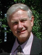
Ph.D. in Radiation Physics from the University of London. Past Secretary-General of the IOMP and Past President of several organizations including the IOMP, IUPESM, IMPCB, AAPM, ACMP, and ABS. Chief Medical Physicist at New York University School of Medicine, Rhode Island Hospital and Brown University, and Karmanos Cancer Institute and Wayne State University, where I am currently Professor Emeritus. Past Editor of Medical Physics World, Medical Physics, Bulletin of the American College of Medical Physics, and the AAPM Quarterly Bulletin. Author or coauthor of about 300 papers in refereed journals, over 50 book chapters and 600 presentations, and author, coauthor, or editor of over 50 books. Major research interests have been bioeffect dose modeling, development of new fractionation and dose-rate regimes, HDR brachytherapy, cervix and breast radiotherapy, radiobiology, radiation carcinogenesis, radiation induced injuries, and radiotherapy physics.
Abstract:
I will be reviewing experiences when helping incoming Vice-President Prof. Larry Lanzl establish Medical Physics World in 1982 and as MPW Editor from 1985-1988, as IOMP Secretary-General from 1988-1994, and as IOMP President from 2000-2003. Memorable events during my terms of office included the establishment of the IOMP Library Program, the Travel Scholarships program for attendance at the World Congress by developing country delegates, the publishing agreement between the IOMP and the IOP, the IOMP/AAPM International Scientific Exchange Program (ISEP), the Donation of New and Used Equipment program, and the awarding of Full Membership in ICSU. Personal memories include helping countries establish their national medical physics organizations so as to apply for IOMP membership, participating in the 1st IOMP/AAPM ISEP course in Pakistan in 1992, and participation in the 1991 USSR medical physics conference when the USSR was dissolving, and the member countries voted to form their own national medical physics societies.
Speaker: Fridtjof Nüsslin, Technical University Munich, Germany
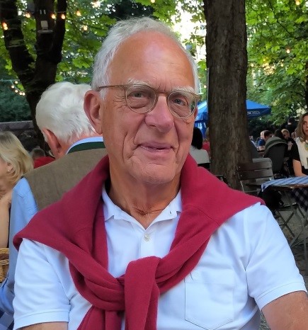
Education & Academic Qualification:
1959 Study of Physics and Medical Sciences at universities of Tübingen and Heidelberg;
1968 Dr.rer.nat., Physics and Physiology, University of Heidelberg; Postdoctoral fellowship at Max-Planck-Institute for Nuclear Physics, Heidelberg;
1979 Qualification university lecturer in Medical Physics;
1984 Professor Medical Physics;
Employment:
1970 Physicist at Radiotherapy Department, Medical School Hannover;
1987-2004 Full professor Medical Physics at the University of Tübingen;
Scientific Activities & Topics of interest:
Radiotherapy Treatment Optimisation, Conformal Radiotherapy (IMRT, IGRT); Dosimetry and Treatment Planning Optimization, Conformal Radiotherapy, Image Guidance, Advanced Technologies (particle beam therapy, laser application in imaging & particle beam therapy), Imaging technology (MRI/MRS, PET, CT); Biological & Molecular Imaging, Biological Modeling, Small Animal Image Guided High Precision Irradiation Technology. Treatment planning, Radiotherapy equipment, Dosimetry; Quality Assurance in Radiotherapy and Radiology; Radiation Protection; Non-Ionizing Radiation Effects; Hyperthermia; Radiological and Nuclear Emergencies. National and International professional matters of Medical Physicists, Medical Physics in developing countries. Education & Training Programmes
Professional Activities:
Since 2004 Professor Biomedical Physics at Technische Universität München
Various positions held in scientific organisations, boards, committees in Germany;
1990-1994 president DGMP
1994-1996 EFOMP Chair Scientific Committee;
1996-1998 EFOMP President.
2000-2006 IOMP Chair International Advisory Board
2006-2009 IOMP Vice President (elect)
2009-2012 IOMP President
2012-2015 IUPESM Vice-President
Honours:
2004 Honorary membership of the Czech Association for Medical Physicists (CAMP)
2005 Honorary membership of the European Federation of Organisations for Medical Physics (EFOMP)
2006 Honorary membership DEGRO (German Society of Radiooncology)
2008 Honorary membership OEGRO (Austrian Society of Radiooncology)
2008 Distinguished Affiliated Professor, Technische Universität München (TUM) and Fellow of the Institute of Advanced Studies, Technische Universität München (TUM-IAS)
2011 Richard-Glocker-Award of the DGMP (German Society of Medical Physics)
2011 Honorary membership SMPS (Saudi Medical Physics Society)
2013 Honorary Fellow IOMP
2021 Honorary Member DGMP
2021 Honorary Fellow IUPESM
Publications:
More than 200 publications in peer reviewed journals
Abstract:
Why medical physics? What is so fascinating in medical physics? Isn´t it just something in between of medicine and physics? Looking back on my professional life, I still have the feeling that hardly any other profession than medical physics covers such a broad spectrum of various disciplines. Indeed, during my whole life as a medical physicist I appreciated so much the many opportunities to witness exciting innovations, new trends and breakthroughs in medical and natural sciences, and to enjoy the challenge of translating these into health care practice. Another track of my professional life was my passion for contributing to the expansion and flourishing of medical physics worldwide. In this context it was a great honour and my personal highlight to have the opportunity to serve our community in several capacities at IOMP from 2000-2015. I am indebted to innumerable colleagues and friends during that time who made it possible to achieve important milestones, such as the expansion of the IOMP-Chapter structure, membership of IUPAP, tighter connection to WHO and IAEA, the approved definition of the “Medical Physicist” as a profession listed at the ILO, and many other advancements. However, there are still some issues we must focus on: the harmonization of education & training worldwide, filling the gap of qualified medical physicists, particularly in the LMI-countries, and opportunities for advancing medical physics science.
25 Oct 2023: Radiopharmaceutical Therapy (RPT)
Wednesday, 25th October 2023 at 12 pm GMT; Duration 1 hour
Organizer: M Mahesh
Moderator: M Mahesh
Speakers: George Sgouros and Ana Kiess
Topic 1: Imaging and Dosimetry in Radiopharmaceutical therapy
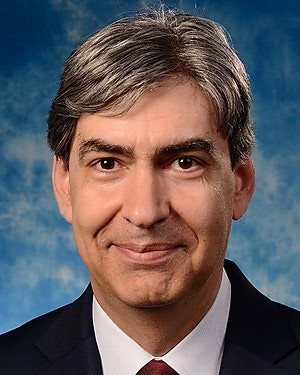
George Sgouros, PhD
Dr. Sgouros is Professor and Director of the Radiological Physics Division in the Department of Radiology at Johns Hopkins University, School of Medicine. He received his PhD from Cornell University, Biophysics Dept, completed his post-doc at Memorial Hospital Medical Physics Dept. He is author on more than 200 peer-reviewed articles, several book chapters and review articles. He is recipient of the SNMMI Saul Hertz Award for outstanding achievements and contributions in radionuclide therapy and a fellow of the American Association of Physicists in Medicine (AAPM). He is a member of the Medical Internal Radionuclide Dose (MIRD) Committee of the Society of Nuclear Medicine and Molecular Imaging (SNMMI), which he chaired 2008-2019. He has chaired a Dosimetry & Radiobiology Panel at a DOE alpha-emitters workshop and also an ICRU report committee for ICRU guidance document No. 96. Dr. Sgouros is a former chair (2015-2017) of the NIH study section on Radiation Therapeutics and Biology (RTB). Dr. Sgouros is also founder and principal of Rapid, a dosimetry and imaging services and software products start-up in support of radiopharmaceutical therapy.
Abstract
Even after they have made it to Phase I clinical trial investigation, 97% of new cancer drugs fail. The majority of these drugs are chosen based on their ability to inhibit cell signaling pathways responsible for maintaining a cancer phenotype. Although this approach has led to dramatic improvements in treatment efficacy for certain cancers, this approach to cancer therapy is more complex than initially appreciated. Radiopharmaceutical therapy (RPT) involves the targeted delivery of radiation to tumor cells or to the tumor microenvironment. Since the radionuclides used in RPT also emit photons, nuclear medicine imaging may be used to measure the pharmacokinetics of the therapeutic agent and estimate tumor and normal organ absorbed doses in individual patients to implement an individualized (precision medicine) treatment planning approach to RPT delivery. This unique feature of RPT, along with its ability to delivery highly potent alpha-particle radiation to targeted cells, is at the heart of what distinguishes RPT compared to other cancer treatments for widespread metastases.
Learning Objectives:
- Understand the mechanism of Radiopharmaceutical therapy.
- Compare and contrast RPT with other cancer therapy modalities.
- Understand the distinction between RPT and Theranostics.
Topic 2: Clinical Radiopharmaceutical Therapy, Dose-Response and Future Directions
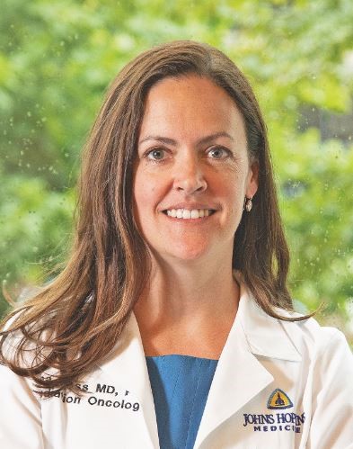
Ana Kiess, MD, PhD
Dr. Ana Kiess’s clinical focus is on the treatment of prostate cancer and head and neck cancers with radiopharmaceutical therapies and external beam radiotherapy. Her research concentrates on the integration of dosimetry, dose-response analyses, and new radiopharmaceutical therapies into the clinic.
Education:
MD; Duke University School of Medicine (2008)
PhD; Biomedical Engineering; Duke University (2008)
Residency:
Radiation Oncology; Memorial Sloan-Kettering Cancer Center (2013)
Recent Publications:
- Wang J, Kiess AP. PSMA-targeted therapy for non-prostate cancers. Front Oncol. 2023 Aug. 13:1220586. doi: 10.3389/fonc.2023.1220586.
- Kiess AP, Hobbs RF, Bednarz B, Knox SJ, Meredith R, Escorcia FE. ASTRO’s framework for radiopharmaceutical therapy curriculum development for trainees. Int J Radiat Oncol Biol Phys. 2022; 113(4):719-726.
- Jia AY, Kashani R, Zaorsky NG, Baumann BC, Michalski J, Zoberi JE, Kiess AP, Spratt DE. Lutetium-177 Prostate-Specific Membrane Antigen Therapy: A Practical Review. Pract Radiat Oncol. 2022; 12(4): 294-299.
- Jia AY, Kiess AP, Li Q, Antonarakis ES. Radiotheranostics in advanced prostate cancer: Current and future directions. Prostate Cancer and Prostatic Diseases. 2023, April: 1-11.
Abstract
Radiopharmaceuticals are rapidly expanding in clinical use and development for prostate cancer and many other tumor types. As in other radiation therapies, there is a dose-response relationship for both tumor and normal tissues, with increasing responses or toxicities at higher absorbed doses. In this webinar, we will discuss these concepts in relation to currently approved radiopharmaceutical therapies (RPTs) and future directions of RPTs. We will also review clinical indications and practical use of currently approved RPTs including [177Lu] Lu-PSMA-617, [177Lu] Lu-DOTATATE, and [223Ra] RaCl2.
Learning Objectives:
- Understand clinical indications and practical use of currently approved radiopharmaceutical therapies
- Discuss concepts of radiation dose and response of tumors and normal organ toxicities
- Explore future directions of clinical radiopharmaceutical therapies.
12 Sep 2023: Physics and Technology for Cancer Care – Meet the IOMP Corporate Members
Tuesday, 12th September 2023 at 12 pm GMT; Duration 1 hour
Organizer: Magdalena Stoeva
Moderator: Ibrahim Duhaini
Speakers: Axel Hoffmann (PTW) & Alexander Pegram (RadFormation)
Topic 1: PTW and VERIQA RT EPID 3D as the Approach to EPID Dosimetry
Axel Hoffmann
Sales Director, PTW, Freiburg, Germany
Education:
09/1969 – 07/1977 Polytecnic Secondary School, Basic Education in Lauta, Germany
09/1977 – 07/1981 Extended Secondary school, Hoyerswerda, Germany
10/1984– 02/1989 Ilmenau Technical University, Ilmenau, Germany. Technical and Biomedical Cybernetics; Biomedical Engineering; Master of Science.
Professional:
02/1989 – 02/1991 National Board for Atomic Safety and Radiation Protection (SAAS), Berlin, Germany. Radiation Measurement in Health Physics; Film Dosimetry.
03/1991 – 08/2013 PTW, Freiburg, Germany, Area Sales Manager (Eastern Europe, Asia, Australia)
09/2013 – Present PTW, Freiburg, Germany, Sales Director, Sales Operation
Abstract
PTW is a world-wide leader in dosimetry. We develop, manufacture, and distribute measurement equipment and software for quality assurance as performed in radiotherapy, diagnostic radiology, and metrology.
PTW is over 100 years old and represents a high level of expertise in our business fields.
VERIQA RT EPID 3D is a new PTW software product for EPID patient treatment plan and delivery verification (pre-treatment and in-vivo). The result is a dose distribution in patient anatomy (based on the back-projection algorithm developed by the Netherlands Cancer Institute). The combination of a Monte Carlo algorithm and the EPID images provide a workflow-efficient and highly accurate dose reconstruction.
The pre-treatment verification does not require a phantom (measurement “in air”). Neither a phantom setup nor a re-planning is necessary.
Topic 2: Optimizing Cancer Care with Efficient QA
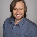 Alexander Pegram
Alexander “Alex” Pegram, DMP, DABR, received his Professional Doctorate in Medical Physics and Master of Science in Medical Physics from Vanderbilt University. His research focused on risk analysis using System Theoretic Procedures (STPA). He also attained certification in Therapeutic Medical Physics from the American Board of Radiology.
At Vanderbilt, Alex was the Chief Medical Physics Resident, before moving to Sanford Health. As the Chief Physicist there, Alex managed a team across two sites and oversaw the purchase, installation, and commissioning of new equipment, including a TrueBeam. In 2020 Alex started at Radformation, where he is now the Product Manager of RadMachine, the company’s cloud-based Radiation Therapy Machine QA Platform. As an affiliate of AAPM, he is part of a working group on “Ask the Expert.”
Abstract
Ensuring the accuracy and reliability of radiation therapy machines is paramount for patient safety and effective cancer treatment in medical physics. In “Optimizing Cancer Care with Efficient QA,” Alex Pegram, DMP, DABR, delves into RadMachine, an innovative solution for Machine Quality Assurance offered by Radformation. RadMachine streamlines the QA process and enhances treatment precision, enabling physicists to perform more efficient, high-quality QA in less time.
The presentation discusses the clinical and scientific aspects of RadMachine, illustrating its seamless integration with data from therapy machines, imaging devices, and ancillary equipment, and more into a consolidated platform. A sophisticated combination of robust automation and a streamlined, cloud-based platform enables RadMachine to efficiently verify and validate machine parameters, minimizing human errors to maximize patient-centric care.
By optimizing resource allocation and time management, the highest standards of safety and quality for cancer care are met. Coupled with additional solutions for workflow automation from start to finish, this presentation will provide insights into the transformative potential of physics-driven technology in delivering exceptional patient care.
29 Aug 2023: IOMP-IUPESM-ISC Joint Webinar: Practical Deep-Dive on ChatGPT
Tuesday, 29th August 2023 at 12 pm GMT; Duration 1 hour
Organizers: Magdalena Stoeva (IOMP,IUPESM) & Zhenya Tsoy (ISC)
Moderator: Magdalena Stoeva
Speaker: Nick Scott

As a senior digital leader for more than 15 years, Nick worked with third sector organizations as diverse as Médicins Sans Frontières, UNISON and the Overseas Development Institute (ODI) to understand their goals; explore the possibilities digital offers for achieving these goals; and map and deliver the changes they needed to succeed. For his work building a digital team at UNISON Nick was listed in the Digital Leaders 100 list in the UK while at ODI his digital strategy was awarded Digital Strategy of the Year at the European Digital Communications Awards.
Since 2021 Nick has been working as a consultant while undertaking an Executive MBA programme at ESADE Business School in Barcelona. Nick helps non profits realize a transformative vision of what they could be in the digital age.
Abstract
The joint IOMP-IUPESM-ISC (International Science Council) webinar will create a safe space for experimentation and explore how the most popular tool – ChatGPT – can be used in our everyday work. The webinar in the form of practical workshop follows the initial workshop organized by the ISC in June 2023.
The webinar is focused on but not limited to the following items:
- An overview of technical capabilities and limitations of ChatGPT.
- A guided live preview of ChatGPT with examples on writing, testing and validating effective prompts.
- What is happening next with new versions of software, such as MS Office, where similar tools will be in-built.
- Moderated Q&A session.
A special focus is given on insights on how to work around technical capabilities and limitations of the tool, what makes for an effective prompt, and other practical questions around the use of ChatGPT.
The webinar is aimed at those working in the scientific organizations, communications colleagues and those with a general interest in using the tool. It would be useful to all levels of users – from those just starting out to discover ChatGPT, to those who are already actively using it.
19 Jul 2023: IOMP-AAPM Webinar
Wednesday, 19th July 2023 at 12 pm GMT; Duration 1 hour
Organizers: Robert Jeraj, Allen Movahed, Claire Park, Robert Jeraj & AAPM Global Research & Scientific Innovation Committee (GRSIC)
Moderator: M. Mahesh (Scientific Committee Chair of the IOMP)
Speakers: Kevin W Eliceiri, Heather Whitney, Robert Finnegan
Title 1: Open Source Computational Imaging of Cellular Microenvironments
Speaker: Kevin W Eliceiri
Kevin Eliceiri is the RRF Walter H. Helmerich Professor of Medical Physics and Biomedical Engineering and Director of the Center for Quantitative Cell Imaging at the University of Wisconsin-Madison. He is also an investigator in the Morgridge Institute for Research. In 2022 he was named a Open hardware Trailblazer by the Open Source Hardware Association and the Sloan Foundation. His research focus involves developing novel optical imaging methods for investigating cellular microenvironment changes in wound healing, and the development of software for multidimensional image analysis. His lab has been contributing lead developers to several open-source imaging software packages including FIJI, ImageJ and μManager. ImageJ was named by Nature as one of the top ten computer codes that changed science. His open hardware instrumentation efforts involve novel forms of polarization, light sheet, laser scanning and multiscale imaging.
Abstract:
A primary interest of the Eliceiri lab is in developing novel imaging techniques that examine the interaction between cells and their microenvironment in space and time. Much of this work has involved the development of multiphoton based and computational approaches to interrogate cells noninvasively deep into intact tissue over time to study metabolism, cell signaling and stromal interactions. This has been particularly useful in studies of cell differentiation and cancer invasion and progression. A major element of this work is the construction of novel open source imaging hardware platforms including custom fluorescent lifetime, spectral imaging systems and multiphoton laser scanning systems. This work entailed new open hardware developments in laser, detection and acquisition technology. Much of our focus in hardware development is at the intersection of hardware and computation. Our past efforts have mainly focused on open source image analysis and visualization (ImageJ), on the computational side, and image acquisition (Micro-Manager) and device management on the hardware side. We are lead developers of the open-source project ImageJ/FIJI and in 2020 we launched the NIH P41 funded “Center for Open Bioimage Analysis” (COBA) to develop and maintain the open-source software CellProfiler and ImageJ while making new deep learning tools and workflow solutions for bioimaging. In 2021 we became the lead development home for Micro-Manager and OpenScan for light microscopy acquisition. We have been leading efforts to build an open source imaging informatics toolkit for light microscopy that spans the complete life cycle of imaging from acquisition, image analysis, data storage, and data dissemination. These tools have included support for novel imaging modalities such as light sheet microscopy, new metadata standards for sharing data and improved image analysis packages such as our work on ImageJ. This work on open source imaging software has enabled novel biological imaging studies and is in use by thousands of labs around the world. We now realize that there is an outstanding, untapped opportunity to advance computational optics, including opportunities for image analysis at runtime, where image processing routines can be run during acquisition to yield new insights and outcomes on data that would not be possible post-acquisition. We are combining our hardware accelerated approaches with our new algorithms to develop “smart” microscopes that can make decisions at runtime and use these decisions to determine where to focus in large tissue arrays.
Title 2: Open source tools from the Medical Imaging and Data Resource Center
Speaker: Heather Whitney
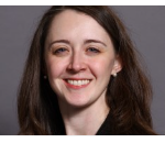
Heather M. Whitney, PhD is a research assistant professor in the Department of Radiology at the University of Chicago. She conducts research in computer-aided diagnosis of breast and ovarian cancer, focusing on the modalities of dynamic contrast-enhanced magnetic resonance imaging and ultrasound. Her primary areas of interest are in artificial intelligence and radiomics across the imaging and classification pipeline, from medical image acquisition to performance evaluation and data harmonization. She also conducts research and collaborates in MIDRC, the Medical Imaging and Data Resource Center. Within MIDRC she works on methods of task-based distributions, interoperability between data enclaves, and monitoring and studying the diversity and representativeness of the MIDRC data commons to foster research in AI and health disparities.
Abstract:
The Medical Imaging and Data Resource Center (MIDRC, midrc.org) is a multi-institutional collaborative initiative driven by the medical imaging community that was initiated in late summer 2020 to help combat the global COVID-19 health emergency and to accelerate the transfer of knowledge and innovation in the COVID-19 pandemic and beyond. MIDRC, funded by the National Institute of Biomedical Imaging and Bioengineering (NIBIB) and hosted at the University of Chicago, is co-led by the American College of Radiology®, the Radiological Society of North America, and the AAPM. The aim of MIDRC is to foster machine learning innovation through data sharing for rapid and flexible collection, analysis, and dissemination of imaging and associated clinical data by providing researchers with unparalleled resources. In this talk, I will discuss the MIDRC open data commons and freely available resources developed by MIDRC investigators, including tools for building patient cohorts, building bias awareness in AI/ML development, and selecting performance metrics, as well as opportunities for contributing data.
Title 3: Unlocking medical imaging with platipy
Speaker: Robert Finnegan
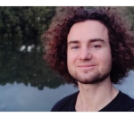
Rob, an Australian physicist, brings extensive experience from geophysics, astrophysics, and medical physics. After completing his PhD from the University of Sydney he continued his work though research fellowships across multiple institutes, where he co-established the platipy library to facilitate accessible, high-quality tools for medical imaging and radiotherapy. Currently, Rob is advancing his skills through clinical medical physics training at the Northern Sydney Cancer Centre.
Abstract:
In the evolving landscape of medical physics, open-source software is becoming increasingly pivotal. This talk will introduce PlatiPy, a Python library designed to revolutionise medical
imaging. PlatiPy offers a comprehensive, extensible, and continually expanding suite of tools, spanning all aspects of processing and analysis such as DICOM conversion, image registration, automatic segmentation, and a powerful visualisation toolkit. This talk will delve into the capabilities of PlatiPy, demonstrating simplify and expand users’ capabilities to use and understand imaging and radiotherapy data.
A standout feature of PlatiPy is the streamlining of common tasks in big data pipelines, a critical component in today’s data-driven medical research. To illustrate this, we will walk through an end-to-end example focusing on cardiac toxicities. This demonstration will highlight how PlatiPy is innovating medical imaging and radiation dose assessment pipelines. Embracing the principles of accessible and open software, PlatiPy encourages collaboration among researchers, medical physicists, and computer scientists worldwide. Are you ready to explore how PlatiPy is helping to unlock medical imaging? Join us!
20 Jun 2023: Radiation Doses and Risk in Imaging – to Know or Neglect?
Tuesday, 20th June 2023 at 12 pm GMT; Duration 1 hour
Organizer: Prof Magdalena Stoeva
Moderator: Prof Arun Chougule
Speakers: Prof Dr Anchali Krisanachinda and Prof Dr Tomas Kron
Title 1: Imaging doses in radiotherapy: to know or neglect?
Speaker: Prof Dr Tomas Kron
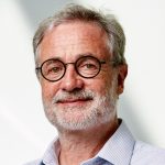
Tomas Kron was born and educated in Germany. After his PhD he migrated to Australia in 1989 where he commenced his career in radiotherapy physics. From 2001 to 2005 he moved to Canada where he worked at the London Regional Cancer Centre on the commissioning of one of the first tomotherapy units. In 2005, Tomas became principal research physicist at Peter MacCallum Cancer Centre in Melbourne, Australia where he now is Director of Physical Sciences. He holds academic appointments at Wollongong, RMIT and Melbourne Universities. Tomas has an interest in education of medical physicists, dosimetry of ionising radiation, image guidance and clinical trials demonstrated by 100 invited conference presentations and 330 papers in refereed journals. He has received many awards over the years including an Order of Australia Medal (2014), Fellowship of IOMP and IUPESM and Life Membership of the TransTasman Radiation Oncology Group (TROG) in 2020.
Abstract:
Radiotherapy is one of the main treatment modalities for cancer patient. It uses target doses in excess of 50 Gy to eradicate tumour cells. In the context of this high dose many people consider dose from imaging procedures that allow for treatment planning and delivery verification to be negligible, a position reflected in the lack of education about imaging dose optimisation for radiotherapy physicists and scant dose reduction methods available on commercial radiotherapy equipment. This presentation argues that this is a mistake not only because the framework of radiation protection urges us to justify and optimise all radiation doses. The number and complexity of imaging procedures is increasing and the volume of the patient irradiated and dose distribution in imaging is fundamentally different form the high dose region in therapy. As such a recently formed task group of ICRP is dealing with imaging dose in radiotherapy. This and experience of managing imaging dose in a large radiotherapy centre will be subject of the presentation.
Title 2: Radiation-induced Cancer Risk in Medical Imaging: To know or Neglect?
Speaker: Prof Dr Anchali Krisanachinda
Anchali Krisanachinda graduated her B.Sc.(Hons) in Physics, M.Sc. (Radiation Physics) from University of London, UK, and Ph.D. (Medical Radiation Physics) from University of Health Science, North Chicago, Ill, USA. She was a Director of Medical Physics Graduate Program, Mahidol University, Bangkok, Thailand. She established Medical Imaging/Medical Physics, graduate programs, M.Sc. and Ph.D. at Chulalongkorn University and became Chairperson of both programs. She established Thai Medical Physicist Society (TMPS) in 2002, started the clinical training of medical physicists in 2007 and obtained Clinically Qualified Medical Physicist, CQMP, in radiation oncology, ROMP in 2009, DRMP in 2012 and NMMP in 2015. Then all 3 branches in medical physics clinical training were started at the same time using AMPLE (Advanced Medical Physics Learning Environment). Medical Physicists in South-East Asia join AMPLE sharing Thai Clinical Supervisors in ROMP and NMMP. The Ministry of Public Health of Thailand approved the medical physics national license in 2022, more than ten years after she requested. Hopefully, in 2023, there will be 300 Thai medical physicist with national license in medical physics. Continue Professional Development in medical physics will be later established. She has received many awards from her Faculty of Medicine and Chulalongkorn University, South-East Asian Federation of Organizations for Medical Physics (SEAFOMP), Asia-Oceania Federation of Organizations of Medical Physics (AFOMP), IOMP and IUPESM.
Abstract:
The risk model, the cancer sites, dose and dose rate effectiveness factor (DDREF) and mathematical models will be described presenting differences among the models. These models take into account parameters such as sex, age-at-exposure, attained age and time since exposure.
Medical Imaging:
Patient dose from a chest X-rays is about 0.1 mSv, whole-body CT scan is about 10 mSv. The effective doses from diagnostic CT procedures may be associated with an increase in the possibility of fatal cancer of approximately 1 chance in 2000. This increase in the possibility of a fatal cancer from radiation can be compared to the natural incidence of fatal cancer in the U.S. population, about 1 chance in 5 (400 chances in 2000). In other words, for any one person the risk of radiation-induced cancer is much smaller than the natural risk of cancer. If the natural risk of a fatal cancer is combined to the estimated risk from a 10 mSv CT scan, the total risk may increase from 400 chances in 2000 to 401 chances in 2000. Nevertheless, this small increase in radiation-associated cancer risk for an individual can become a public health concern if large numbers of people undergo increased numbers of CT screening procedures of uncertain benefit.
12 May 2023: Radiogenomics/Radiomics-Guided Personalized Radiation Therapy: Current Status, Challenges, and Opportunities
Friday, 12th May 2023 at 12 pm GMT; Duration 1 hour
Moderator: Prof Arun Chougule
Speaker: Dr. V. Subramani
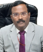
Dr. V. Subramani, PhD is currently working as Assistant Professor of Radiation Oncology (Medical Physics) and Heading Medical Physics in the Department of Radiation Oncology at All India Institute of Medical Scien
ces, New Delhi. He is having more than 26 years of experience in in radiation oncology medical physics. He has publication of around 50 scientific research articles as author and co-authors in the field of radiation oncology. Also He has delivered around 70 invited guest lectures, chaired several scientific sessions and participated in the debates and panel discussions in the national and international professional organization’s annual and periodic scientific meetings for the last ten years in the field of advanced radiotherapy and medical physics. He is currently national secretary of Association of Medical Physicists of India (AMPI), IOMP Education and Training Committee member and also serves as Chief Editor of Asia-Oceania Federations of Organization for Medical Physics Newsletter.
Abstract:
The success of a cancer treatment depends on the ability to deliver the right treatment to right patient to a right dose at right time, which is an ultimate goal of precision medicine or precision oncology. However, currently patients undergoing radiotherapy are treated using uniform dose constraints specific to the tumor and surrounding normal tissues. This population based one-size-fit-all approach results in significant adverse effects and suboptimal tumor control. Another concern with current approach is that two patients with nearly identical dose distributions can have substantially different acute and chronic morbidities leading to poor quality of life. Therefore, there is a need to develop an approach to overcome this limitation of current standard of care in oncology. Due to recent advances in biological and quantitive imaging, image processing analysis, computational power, cancer biology, biotechnology and genomics, there are tremendous growths and large amount of data is available for each individual patient.
The radiogenomics is an emerging field in precision radiation oncology. “Radiogenomics” has two meanings: “the study of genetic variation associated with response to radiation (Radiation Genomics) and “the correlation between cancer imaging features and gene expression (Imaging Genomics). Radiogenomics is a combination of both radiomics and genomics biomarkers, which is useful in guiding and personalizing treatment prescription and adaptation when changes occurring during the course of therapy. This is termed as Radiogenomics-Guided personalized radiation therapy. Both radiomics and radiogenomics biomarkers can be used to evaluate disease characteristics or correlate with relevant clinical outcome such as patient prognosis and treatment response. The common goal of discovering useful diagnostics, prognostics or predictive biomarkers to improve clinical decision making and ultimately enable the practice of precision and personalized medicine.
The presentation will address about the medical physics aspects of quantitative imaging biomarker, radiomics/radiogenomics model development, radiomics-guided radiotherapy using radiomics-target volume, radiomics-knowledge based treatment and also gnomically-guided radiotherapy including genomic-adjusted radiation dose and radiation sensitivity signature and models for personalized radiotherapy.
28 Apr 2023: IMPW 2023 Webinar: Upright Radiotherapy: Challenges and Opportunities
View Recording
Friday, 28th April 2023 at 12 pm GMT; Duration 1 hour
Organizer: Eva Bezak, IOMP
Moderator: M Mahesh, IOMP
Speaker: Tracy Underwood, PhD

Dr Tracy Underwood is a Senior Physicist at Leo Cancer Care, an innovative young company who are developing medical devices for upright radiotherapy. She is also a UKRI Future Leaders Fellow and an Honorary Senior Research Fellow at University College London. As a radiotherapy researcher she has worked at some of the most innovative clinics and academic departments worldwide, including Massachusetts General Hospital / Harvard Medical School in Boston, IUCT Oncopole in Toulouse, the Christie NHS Foundation Trust, the University of Manchester, and the University of Oxford. She is passionate about medical physics education and is part of the editorial board of “The Encyclopaedia of Medical Physics”, which is published as an open resource at http://www.emitel2.eu/, as well as in hardcopy by CRC Press. Tracy was awarded the 2015 IMechE JRI: Best Medical Engineering PhD Prize and the 2017 Early Career Academic Prize from the UK Institute of Physics and Engineering in Medicine. She likes to spend her free time on the beach or in the forest with her husband and two young kids.
Abstract:
Treatments which combine fixed radiation beams with upright, rotating patient positioning systems have been investigated since the advent of radiotherapy. However, recently there has been an upsurge of interest in this topic, as evidenced by new academic publications and commercial systems. In Heidelberg, one of the only carbon ion gantries in the world weighs ~600 tonnes. For proton therapy, gantries typically weigh ~100-200 tonnes. Even conventional photon gantries weigh ~5 tonnes: around the weight of an adult elephant. Eliminating the gantry could vastly reduce the radiation shielding required and the 3D footprint of treatment bunkers. It could also reduce the complexity of beam delivery equipment, leading to reduced maintenance, easier upgrades, and lower expertise barriers. These factors can bring down treatment room costs, not just for heavy ion / proton radiotherapy, but also for conventional photon radiotherapy where global access is unacceptably low. There may be tumour sites where upright radiotherapy is better medicine (e.g., lung, liver). For certain patients (e.g., those with head and neck cancers), upright positioning is also likely to prove more comfortable. Against the advantages of upright radiotherapy, we need to weigh the practical challenges of re-implementing the clinical workflow. Questions also remain over immobilisation and how treatment plan quality and radiotherapy effectiveness will vary with body position. Given the decades of enhancements across supine radiotherapy, how committed are we to the traditional paradigm? Here we will explore the published evidence, challenges and opportunities associated with upright radiotherapy.
27 Apr 2023: IMPW 2023 Webinar: Leadership in Medical Physics
Thursday, 27th April 2023 at 12 pm GMT; Duration 1 hour
Organizer: Eva Bezak, IOMP, Simone Kodlulovich Renha, IOMP
Moderator: Simone Kodlulovich Renha, IOMP
Speaker: Colin Orton, PhD
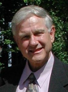
Dr. Orton graduated with a Ph.D. in Radiation Physics from the University of London, England in 1966. He has worked as Director of Medical Physics at New York University School of Medicine (Assistant Professor 1966-1975), at Rhode Island Hospital and Brown University (Associate Professor 1975-1981), and at the Detroit Medical Center/Karmanos Cancer Institute and Wayne State University (Professor 1981-2003), where he is currently Professor Emeritus in the School of Medicine. While at Wayne State he directed of one of the first accredited medical physics graduate programs, with over 150 M.S. and Ph.D. graduates. His major research interests have been bioeffect dose modeling, development of new fractionation and dose-rate regimes, HDR brachytherapy, cervix and breast radiotherapy, radiobiology, radiation carcinogenesis, radiation induced injuries, and radiotherapy physics, and he has taught courses in radiation biology annually to residents, therapists, and physicists. He has served as President of the American Association of Physicists in Medicine (AAPM), Chairman of the American College of Medical Physics (ACMP), President of the International Organization for Medical Physics, the American Brachytherapy Society (ABS), the International Union for Physical and Engineering Sciences in Medicine (IUPESM), and the International Medical Physics Certification Board. Dr. Orton has published over 20 books, over 290 papers, made over 600 presentations, and has received the major awards of the AAPM, ACMP, ABS, IOMP and IUPESM.
Abstract:
Many medical physicists would like to become leaders, but many will be just as happy working all their lives making valuable contributions to the health and safety of their patients without taking on the added responsibilities of leadership. This talk will review the different types of leadership available to medical physicists, what makes you a “leader”, and what opportunities are there for you to reach your leadership potential.
Leadership can take several forms for medical physicists, such as:
- Chief Medical Physicist
- Chief Clinical Physicist
- Radiation Safety Officer
- Director of an educational program
- Director of a residency training program
- Officer in a regional, national, or international medical physics organization
In this talk we will discuss a variety of topics, including:
- Can anyone be a leader or do leaders need to have special traits (interests, skills, motivation, personality, commitment…)?
- Can leadership be taught and, if so, should a teaching program include “leadership” in the curriculum?
- Should there be leadership mentorship programs?
- How can you work to become a leader?
26 Apr 2023: IMPW 2023 Webinar: Cumulative Dose: What, Why, When, How, and How Much?
Wednesday, 26th April 2023 at 12 pm GMT; Duration 1 hour
Organizer: John Damilakis, IOMP
Moderator: John Damilakis, IOMP
Speaker: Madan Rehani, PhD
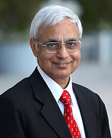
Dr. Madan Rehani is Director, Global Outreach for Radiation Protection at the Massachusetts General Hospital, Boston. He was President, IOMP (2018- 2022) and is currently President IUPESM. He is a retired Professor of Medical Physics and also retired from the IAEA where he worked for 11 years. He was Head of the WHO Collaborating Center on Imaging Technology and Radiation Protection in India. Dr. Rehani is an Emeritus Member, International Commission on Radiological Protection (ICRP), having been active member for 24 years. He is author of 9 Annals of ICRP, 4 of which as Chair of the TG. He is Senior editor Br J Radiology and Assoc Editor, Eur J Medical Physics. He has more than 180 publications, has written 40 chapters in Books and edited 5 books. Besides medical physics & radiology journals, he has published papers in clinical journals e.g. JAMA Intern Med, Br Med J, Eur Heart J, Cardiovascular Imaging, Am J Gastroenterol, Circulation J, The Lancet.
Abstract:
Despite criticism, there is no way out from cumulative effective dose (CED) when several organs are involved, and we have to focus on the patient’s radiation safety in addition to the technique dose. One cannot close eyes to stochastic risks when patients are receiving cumulative doses in three digits of mGy of organ doses or three digits of mSv of CED in a short period of a few years. Scientists involved in studying radiation effects inform us that we have to sum doses from imaging exams performed at different times and they do not have gap correction factors for stochastic effects. Studies published in the last 3 years have brought new results, never before known. They have opened a new era where every one of us has the potential to contribute to enhancing patient radiation safety and the manufacturers have to do much more. The talk will deal with what lies ahead, which is more powerful than controversies.
25 Apr 2023: IMPW 2023 Webinar: Micro-X CNT Emitters, X-ray Tubes, and Unique Imaging Applications
Tuesday, 25th April 2023 at 12 pm GMT; Duration 1 hour
Organizer: Eva Bezak, IOMP
Moderator: Eva Bezak, IOMP
Speaker: Brian Gonzales, PhD
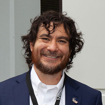
Brian is the Chief Scientist of Micro-X and the CEO of Micro-X Inc, the US subsidiary. Brian has a Ph.D. in biomedical engineering from UNC and NC State University and has published in multiple peer-reviewed scientific journals, focused on X-ray Computed Tomography using carbon nanotube (CNT) X-ray technology. Brian has worked with carbon nanotube X-ray technology from the early research at UNC, then lead early development of the technology at XinRay Systems, and then assisting in the transition to full commercialization of the technology at Micro-X in partnership with Flinders University and the University of Melbourne in Australia.
Abstract:
Micro-X has successfully developed the world’s first carbon nanotube (CNT) based x-ray tube for medical applications. This unique technology achieves both the high x-ray current, and the stable x-ray output required for safe and effective medical imaging. The performance of Micro-X CNT tubes is achieved through two patented design features of the CNT emitter that differentiate the emitter for other field-emission emitters. In this presentation, Micro-X will present an overview of the emitter design and share emitter data demonstrating the high current and stable performance.
CNT x-rays are smaller and simpler compared to conventional x-ray tubes. CNT x-ray tubes are controlled by direct electronic voltage instead of the indirect thermionic control of conventional x-ray. These differences enable new imaging systems to be developed. In this presentation, Micro-X will overview the four different products Micro-X has designed taking advantage of the advantages of the CNT x-ray. This includes a lightweight mobile digital x-ray imaging system to bring x-ray directly to a patient bedside, an x-ray camera that creates two-dimensional x-ray backscatter images with the x-ray source and detector on a single side, a compact lightweight CT for early diagnosis of stroke, and a reimaged airport checkpoint based around a miniaturized CT baggage scanner.
24 Apr 2023: IMPW 203 Webinar: Radiation Protection When Imaging Pregnant Patients: An ICRP Perspective
Monday, 24th April 2023 at 12 pm GMT; Duration 1 hour
Organizer: John Damilakis, IOMP
Moderator: John Damilakis, IOMP
Speaker: Kimberly Applegate, MD. MS
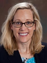
Dr. Kimberly Applegate is a retired professor of radiology and pediatrics from the University of Kentucky in Lexington.Kimberly is a member of the ICRP Main Commission of the International Commission for Radiological Protection (ICRP) as chair of Committee 3, focusing on radiation protection in medicine and also the National Council on Radiation Protection and Measurements (NCRP). She is active on multiple ICRP task groups including TG 108, 109, 113, 121, 124, and 126 and NCRP subcommittees—one on patient shielding in medical imaging and another on the stratification of equipment use and training for fluoroscopy. To improve safe and effective imaging care of children worldwide, Kimberly has been active in the Image Gently Campaign (now the Alliance) Steering Committee from its beginning in 2007.
Abstract:
The radiological protection community in medicine has long recognized the relative radiosensitivity of pregnant women and the fetus compared to the average adult patient. There are a number of serious clinical conditions, including new cancer diagnoses, that occur during pregnancy and that require imaging procedures and radiation therapy that will be described. Further, there is continued evolution of our scientific understanding of radiation health effects, of societal values, and imaging protocols allow for improved radiological protection as long as local education/training and communication are provided for all stakeholders. However, there are varying levels of guidance, imaging protocols, and shared decision-making with women and their families. This webinar will provide an update on the ICRP work regarding this topic.
8 Mar 2023: Women in Medical Physics (on the occasion of the International Women’s Day)
Wednesday, 8th March 2023 at 12 pm GMT; Duration 1 hour
Organizer: Magdalena Stoeva and Eva Bezak
Moderator: Loredana Marcu
Title 1: Improving Women Health: the needs of medical imaging and the IAEA perspective
Speaker: Virginia Tsapaki, PhD

Medical Physicist (Diagnostic Radiology), Dosimetry and Medical Radiation Physics Section, Division of Human Health, Department of Nuclear Applications, International Atomic Energy Agency (IAEA)
Medical Physicist specialised in radiology at the DMRP Section, Division of Human Health, IAEA since August 2019. Since then, involved in multiple regional, national training courses, as well as various missions and activities related to human health. Scientific Secretary of 3 IAAEA publicaitons and contributor to 3 other related guidance documents. From 2004 until 2019, an IAEA expert sent in several missions, analysing data and publishing scientific papers from various IAEA surveys. Clinical experience for approximately 30 years with more than 150 publications in national/international journals/conference proceedings and more than 200 presentations/posters in national/international conferences. Participated in multiple European projects such as the Clinical Diagnostic Reference Levels, ENEN+ project, Basic Safety Standards Transposition project, ENETRAP III, Paediatric Diagnostic Reference Levels, EUTEMPE-RX, EMAN, SENTINEL, DIMOND II/III research projects. Served as a chair or member of committees in international organizations (AAPM, IOMP, EFOMP, ESR, EURAMED). Chair of Organizing committee of 2 European conferences (2016, 2018).
Abstract:
The International Atomic Energy Agency (IAEA) assists Member States in nuclear sciences and applications to benefit human health and to ensure safe, high quality and effective medical uses of radiation. The overall goal of the IAEA Human Health programme is to build capacity and transfer technology for the prevention, diagnosis and treatment of diseases in radiation medicine according to best practices. Various activities to support the above efforts, relevant to improving women health are: a) Guidance documents, training and professional matters; b) Coordinated Research Activities; c) Development of training resources; and d) Clinical audit programmes. All these will be presented in more detail during the session.
Title 2: Pioneer women in medical physics from the Middle East
Speaker: Huda Al Naemi, PhD

Executive Director, Hamad Medical Corporation, A/Professor, Weil Cornel Medicine, Qatar
Dr. Huda has been working in Hamad Medical Corporation for almost 3 decades and she is the Executive Director of Occupational Health and Safety Department since 2006. Dr. Al Naemi represents Qatar in a number of international and global organizations such as; International Atomic Energy Agency (IAEA), IPEM and World Health Organization (WHO) and implement some of their projects at the national level. Dr. Al Naemi is an active member in many international organizations such as, European Society of Radiology(ESR), American Organization of Physicists in Medicine (AAPM), IOMP, Institute of Physics and Engineering in Medicine (IPEM) and, Gulf Nuclear medicine association.
In 2019 Dr. AlNaemi was appointed as Assistant professor of Medical Biophysics Research in Radiology at Weil Cornel Medicine (WCM). Dr. AlNaemi’s role in WCM is to collaborate in education and training for medical students and to conduct research at the cutting edge of knowledge to provide the highest quality of care to the community. Dr. AlNaemi has collaborated on several research and educational projects including some funded by the Qatar National Research Fund (QNRF) and International Atomic Energy Agency (IAEA). These have led to several peer-reviewed publications, this research focused on radiation dose optimization in imaging modalities for patients including pregnant women and pediatrics. The two projects funded by QNRF were in collaboration with Massachusetts General Hospital and Harvard Medical School USA and Geneva University Hospital Switzerland.
Dr. AlNaemi was elected president of the Middle East Federation of Organization of Medical Physics (MEFOMP) for the term 2018 – 2022 and she is also the president of the Qatar Medical Physics Society (QaMPS) since 2018. As MEFOMP president Dr. AlNaemi led the MEFOMP efforts for the publication of a chapter in a published book on “Medical Physics during the COVID-19 Pandemic. In 2019 Dr. Al Naemi was awarded the Institute of Physics and Engineering in Medicine (IPEM) The Healthcare Gold Medal. In 2017 she was awarded the State Encouragement Award for Medical Sciences Category, Doha Qatar. In 2022 Dr. Huda received an appreciation from IOMP for her strenuous efforts in her work as president of MEFOMP from 2018-2022. In 2022 Dr. Al Naemi has been elected as a regular member of the IDMP, one of the IOMP committees.
Abstract:
Main Objectives
To Believe in ourselves as women and face the challenges
To do the maximum energy in work and giving
To be always optimistic and believe of better future.
Middle Eastern women have a very limited opportunity when it comes to professional career and they are thought to be better off staying at home taking care of the household. Yes indeed, such fact existed in the old days. It has rooted from the very own families of the Arabs and continued in primary school where girls and boys studies separately. There is an opportunity then for women, but it was limited only to teaching and nursing profession. But gone are those days; opportunities have opened for women. Slowly, the colleges started admitting women in the so-called “men field” especially in the healthcare industry.
Eventually, I started work in healthcare as senior radiation physicist then became the Executive Director of Occupational Health and Safety Department where I direct various health and safety sections including the Radiation Safety Section which was later named as Medical Physics Section. Being the only Medical Physicist in Qatar then, I represented the country internationally and had the opportunity to work with various international organizations such as WHO, IAEA, UNEP to name a few, and has implemented some of their projects at the national level. It has always been my aspiration to further the advancement of the medical physics profession locally and regionally. I established the Qatar Society of Medical Physics (QSMP) and co-founded the Middle East Federation of Medical Physics (MEFOMP) which I also became the President in 2018-2021.
Title 3: Wearing more than one hat – is this the new fashion trend for women in medical physics?
Speaker: Iuliana Toma-Dasu, PhD

Medical Radiation Physics Division, Stockholm University and Karolinska Institutet, Cancer Center Karolinska, 171 76 Stockholm
Iuliana Toma-Dasu is Professor in Medical Radiation Physics and the Head of the Medical Radiation Physics division at the Department of Physics, Stockholm University, affiliated to the Department of Oncology and Pathology at Karolinska Institutet in Stockholm, Sweden, and the Editor in Chief of Physica Medica – European Journal of Medical Physics.
Iuliana Toma-Dasu studied Medical Physics at Umeå University, Sweden, where she also became a certified medical physicist and received a Ph.D. degree. In parallel with her involvement in the educational program for the medical physicists run at Stockholm University, her main research interests focus on biologically optimised adaptive radiation therapy, including particle therapy, modelling the tumour microenvironment and the risks from radiotherapy.
Abstract:
Achieving gender equality has been one of the objectives of the medical physicists’ community in many European countries, as well as in many other places around the world. This objective has been partially reached, nowadays women in the medical physics being much less underrepresented than in other physics fields. Becoming a medical physicist and performing clinical duties is, therefore, possible for many women due to the various strategies developed and implemented at institutional, national or international levels within the professional associations under the IOMP umbrella. Reaching, however, higher positions in the hierarchy on either clinical or academic side in medical physics is still a considerable challenge for women compared to men. The question therefore is: how many hats should a woman wear, how many accolades should she receive, how many merits should she prove, to be regarded as successful in medical physics and accede to a leading position? This talk would attempt to answer some of these questions while highlighting the importance of successful women in medical physics to share their experience to inspire others and help the policy makers develop the most successful strategies to continue the work towards offering the same opportunities to all genders in the medical physics field.
7 Feb 2023: IOMP Webinar: Carbon-ion Radiotherapy: Current Status and Future Perspective
Tuesday, 7th February 2023 at 12 pm GMT; Duration 1 hour
Organizer: Eva Bezak, IOMP
Speaker: Taku Inaniwa, National Institute for Quantum Science and Technology, Japan
Taku Inaniwa, Ph.D, is a group leader of treatment beam research group at the Institute for Quantum Medical Science, National Institutes for Quantum Science and Technology (QST) in Japan. His research focuses on developing dose calculation algorithms and biological models used for charged-particle therapy treatment planning. He has contributed more than 100 peer reviewed publications. He is a member of international scientific advisory board of Physics in Medicine and Biology. For his works, he has received several national and international awards. From April 2022, he has concurrently served as a guest professor at Division of Health Science, Graduate School of Medicine, Osaka University.
Abstract:
Charged-particle therapy with carbon ions (C-ion RT) has attracted growing interest due to their advantageous physical and biological characteristics. So far more than 40,000 patients have been treated worldwide. During the past three decades, C-ion RT has made remarkable progress in clinical and technological aspects. In my presentation, I would like to introduce physics, dosimetry, radiation biology, carbon RBE, and some clinical results of C-ion RT.
23 Jan 2023: Safety Blinded or Safety Minded – Don’t Learn Safety by Accident
Monday, 23rd January 2023 at 12 pm GMT; Duration 1 hour
Speaker: Chris Trauernicht, President of FAMPO

Chris Trauernicht is the head of the medical physics division at Tygerberg Hospital in Cape Town, South Africa, as well as an associate professor at Stellenbosch University. He is the current president of the Federation of African Medical Physics Organizations (FAMPO) and currently serves on the IOMP Accreditation Board.
Chris has north of 150 congress contributions and 30 papers, and he is a member of the editorial boards of “Advances in Radiation Oncology” and the “South African Journal of Oncology”.
He serves as an assessor for the Health Professions Council of South Africa and has acted as examiner and convenor for the Colleges of Medicine of South Africa. The IAEA has appointed Chris as an expert on numerous occasions, including on the implementation of the Basic Safety Series for medical professionals, on the prevention of accidents and incidents in radiotherapy, and most recently on a regional training course to train the trainers in radiation safety culture (hence the idea for this talk).
Chris was the recipient of the 2020 “International Day of Medical Physics” award for his services to medical physics in the FAMPO region.
Abstract:
Should have, would have, could have… many healthcare professionals may be familiar with that sinking feeling when a preventable incident has slipped through the safety net and reached the patient. The incorporation of safety culture in healthcare can help prevent many adverse incidents and events.
Safety culture can be defined as “the assembly of characteristics and attitudes in organizations and individuals which establishes that, as an overriding priority, protection and safety issues receive the attention warranted by their significance”. In fact, the Bonn Call for Action specifically proposed the strengthening of radiation safety culture as one of its ten main actions.
In 2021, the International Atomic Energy Agency published a booklet on safety culture traits and proposed ten traits – patterns of behaviour or thinking – that encourage the prioritization of safety.
The implementation of these traits is free, yet many systems fail to apply these characteristics effectively. In this talk a brief overview of the ten traits is given. The application of these will improve your institution’s safety culture.

