

🎓
Download your CPD certificate
Only participants who attended a webinar in full are eligible for the certificate.
🕐 To convert webinar times to your local time zone, use the Greenwich Mean Time converter.
🎓 For certificate queries, visit the certificates page.
📢 IOMP School Webinars run approximately monthly — watch this page and IOMP social media for upcoming announcements.
▶ Upcoming
📋 Registration Open — Next Webinar
Date & time
Thu 28 January 2027 · 12:00 GMT
Duration
1 hour
Webinar Overview
Photon-counting computed tomography (PCCT) represents a new paradigm in clinical CT imaging. Unlike conventional energy-integrating detectors (EIDs), photon-counting detectors directly convert individual x-ray photons into electrical signals while simultaneously measuring photon energy. This approach enables smaller detector elements, improved spatial resolution, reduced electronic noise, and intrinsic spectral imaging. The technology offers opportunities for improved visualisation of fine anatomical structures while potentially improving dose efficiency. As with dual-energy CT (DECT), the spectral information acquired with every scan allows material decomposition, virtual monoenergetic imaging, and quantitative characterisation of tissue composition, without requiring a dedicated dual-energy acquisition. Current clinical applications demonstrate particular promise in cardiovascular, pulmonary, musculoskeletal, oncologic, and neuroimaging. This presentation reviews the physical principles underlying PCCT, presents its clinical application in neuroradiology, discusses new emerging technologies in PCCT, and the overall impact on the field of clinical CT imaging.
Learning objectives
- Understand the technology of PCCT.
- Understand the differences between PCCT detectors and EID detectors.
- Recognise the advantages of PCCT detectors over EID.
- Understand the clinical application and advantages of PCCT compared to EID in neuroradiology.
- Learn about updates on PCCT technologies approved for clinical use and those in the pipeline.
Talk 1
Physics of PCCT
Prof. Sameer Tipnis, PhD — Professor of Radiology & Chief of Medical Physics, Department of Radiology and Radiological Science, Medical University of South Carolina (MUSC), USA
▼ Speaker biography
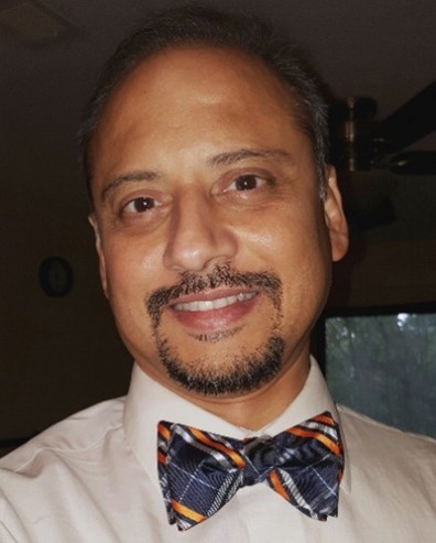
Dr. Sameer Tipnis is a Professor of Radiology & Chief of Medical Physics in the Department of Radiology and Radiological Science at the Medical University of South Carolina, where he provides guidance on optimising imaging protocols in CT, Nuclear Medicine, and radiation safety. He teaches radiology residents, cardiology fellows and medical students about the physics of radiological imaging. He is board-certified by the American Board of Radiology in Diagnostic Medical Physics and Nuclear Medical Physics, serves as ABR trustee for Diagnostic Medical Physics and as an oral examiner for ABR’s Diagnostic and Nuclear Medicine certifying exams. He is a Fellow of the AAPM and chairs task group TG361 on the recommended use and management of protective garments. He is a member of AAPM sub-committees on radiation protection, radiology resident curriculum, and cardiology fellows’ curriculum, and is currently co-Editor-in-Chief of Medical Physics International, a publication of IOMP, and a faculty member at the biennially organised College on Medical Physics at the ICTP, Trieste, Italy. He has published over 50 peer-reviewed articles related to radiological imaging and the development of scintillators.
Talk 2
PCCT in Neuroradiology
Prof. Maria Vittoria Spampinato, MD — Professor of Radiology, Division Director of Neuroradiology, and Vice Chair for Faculty Development, Medical University of South Carolina (MUSC), Charleston, SC, USA
▼ Speaker biography
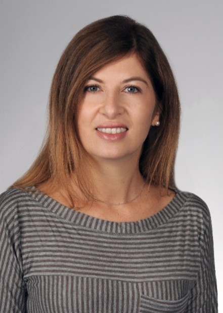
Dr. Maria Vittoria Spampinato is Professor of Radiology, Division Director of Neuroradiology, and Vice Chair for Faculty Development at the Medical University of South Carolina (MUSC), Charleston, SC, USA. She completed her radiology training in Italy, followed by a research fellowship at the National Institutes of Health and a neuroradiology fellowship at the University of North Carolina. Her research has focused on advanced neuroimaging techniques and their clinical applications, including quantitative MRI, diffusion and perfusion imaging, neurodegenerative diseases, and cerebrovascular disorders. More recently, her work has expanded to photon-counting detector CT, with particular emphasis on neurovascular imaging, ultra-high-resolution CT angiography, image quality optimisation, and advanced reconstruction techniques. She has authored more than 80 peer-reviewed publications and has extensive experience leading multidisciplinary initiatives to optimise imaging protocols and integrate emerging technologies into clinical practice. She currently co-leads the Neuroimaging Strategic Area within MUSC’s Neuroscience and Brain Health Institute.
Talk 3
New Technology in PCCT and Overview
Prof. M. Mahesh, MS, PhD — Professor of Radiology, Johns Hopkins University School of Medicine; Professor of Environmental Health and Engineering, Johns Hopkins Bloomberg School of Public Health; Vice-President, IOMP
▼ Speaker biography

M. Mahesh, MS, PhD, is Professor of Radiology at the Johns Hopkins University School of Medicine, Baltimore, MD, USA, and Professor of Environmental Health and Engineering at the Johns Hopkins Bloomberg School of Public Health. He previously served as Chief Physicist for the Johns Hopkins Hospital for over 29 years. He is ABR board-certified, with research interests in medical imaging, artificial intelligence, and radiation risk communication, strongly striving to enhance radiation risk communication with patients, public and media. Dr. Mahesh is an elected member of the ICRP Committee 3 (Medicine) and NCRP; subject-matter expert to the IAEA and UNSCEAR; and Associate Editor of the Journal of the American College of Radiology (JACR). He served as 2nd Vice President of RSNA (2012), and recently as President (2025) and Board Chair (2026) of the AAPM. He has also served on the ACR Board of Chancellors (2018–2024) and the SCCT Board (2015–2018). Dr. Mahesh is currently Vice-President of IOMP.
Past webinars 2027 — recordings available
No past webinars yet in 2027 — recordings will appear here after each live session.
Looking for earlier recordings? See 2026 past webinars.